 Machines & equipment
 Eyecare instruments
 Consumable products
 Other products


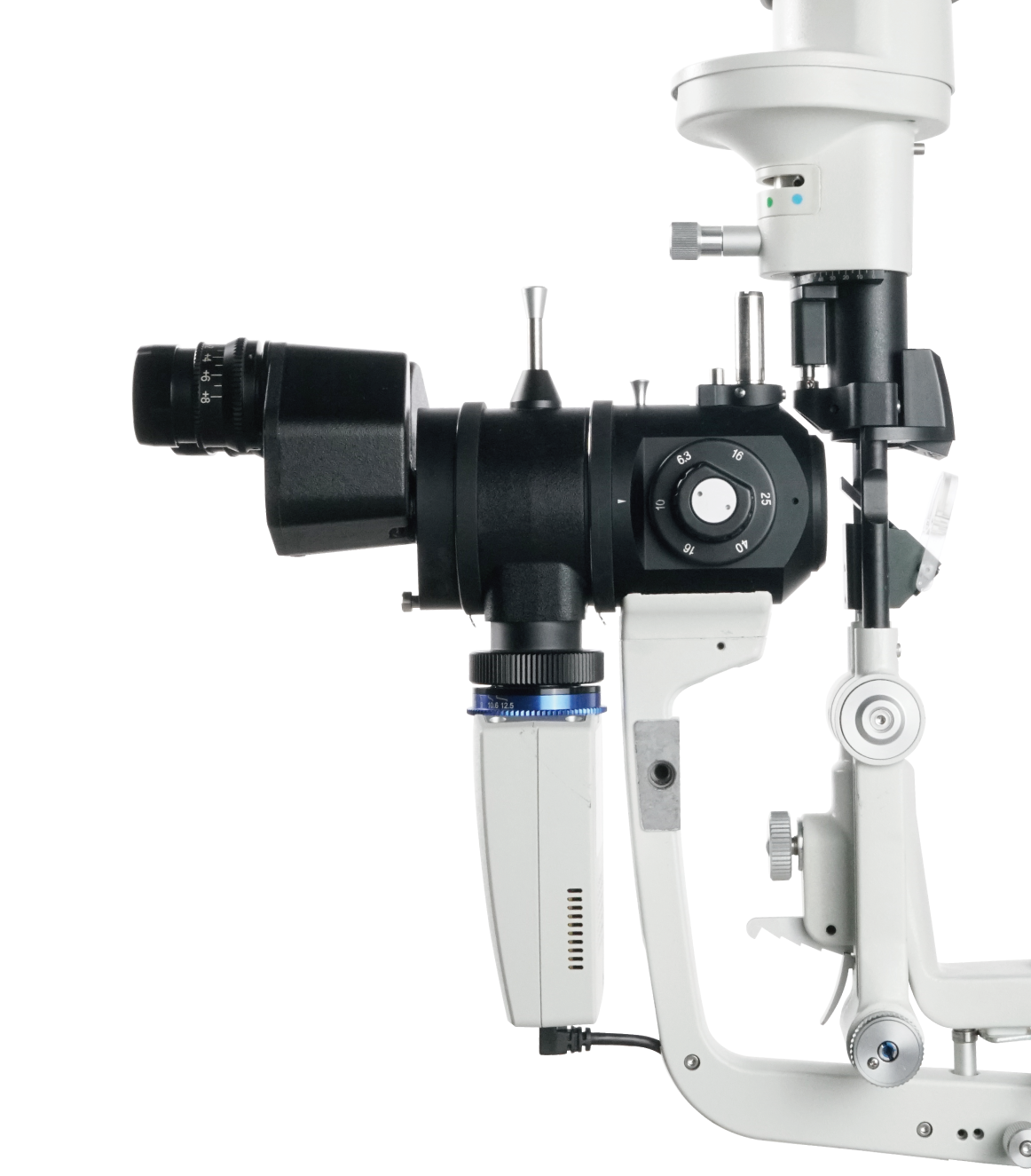
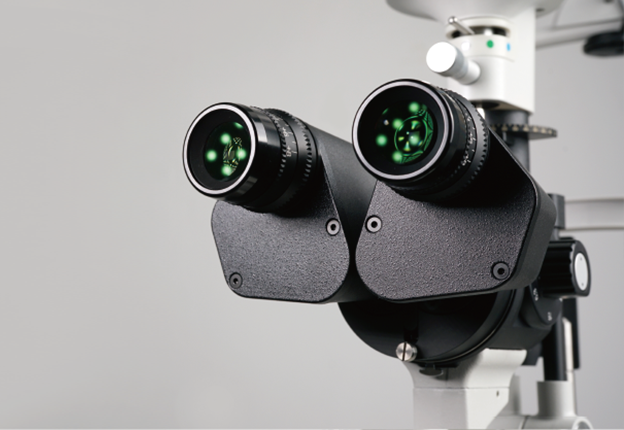
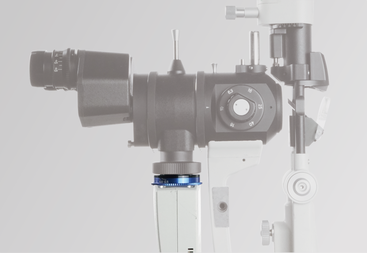
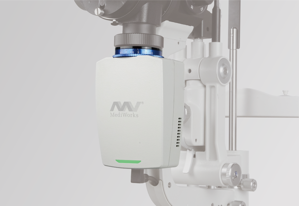
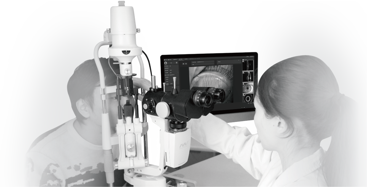
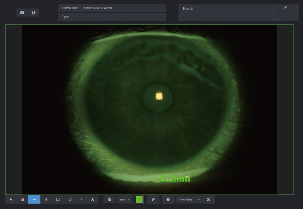
Slit Lamp Microscope – MediWorks – S390L (Firefly)
High sensitivity. The slit is still clear and sharp under weak light.
Wide dynamic range. Iris and sclera images are clearly presented simultaneously with more realistic and evenly distributed colour.
Simple Design + Simple Operation
The design of the ophthalmic slit lamp S390L(Firefly) was inspired by the shape of the firefly. The smart design largely saves space for clinicians compared to other bulky camera systems. We have preset many camera parameters so the user does not need to adjust settings before using the device. The user can operate the machine immediately once the installation has been finished. The device has the following automatic functions for photo shooting and processing when equipped with our Mediview software:
Wide Dynamic Range
Meibomian Glands Examination
Auto Exposure
Auto Gain
Auto White Balance
Auto OD/OS Indicator
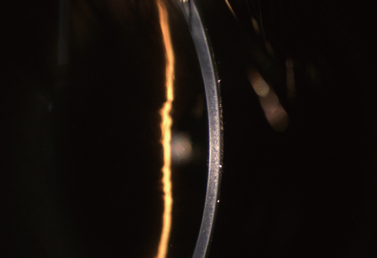
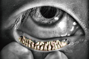
High Sensitivity
The slit is still clear and sharp under weak light.
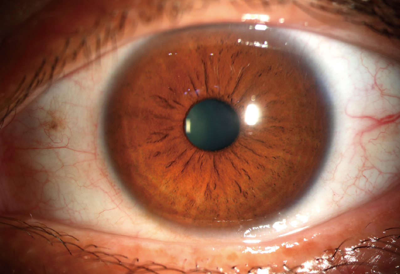
Wide Dynamic Range
Iris and sclera images are simultaneously clearly presented with more realistic and evenly distributed color.
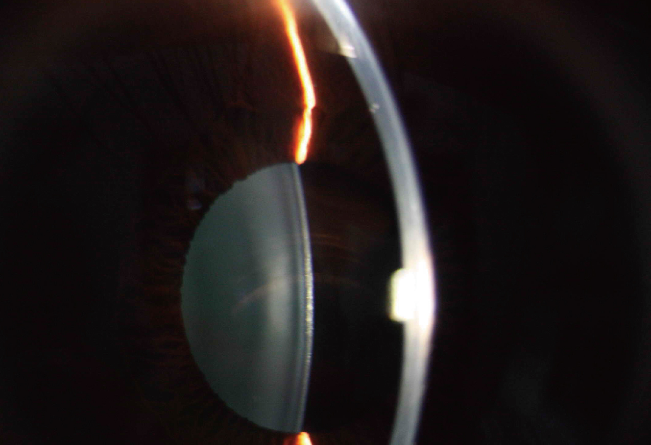
Lens
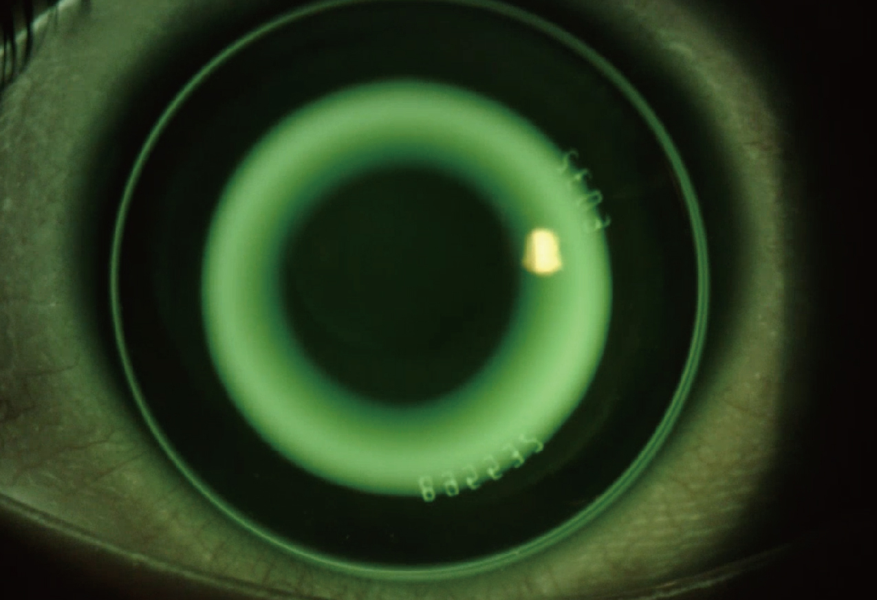
Orthokeratology Lens Fitting
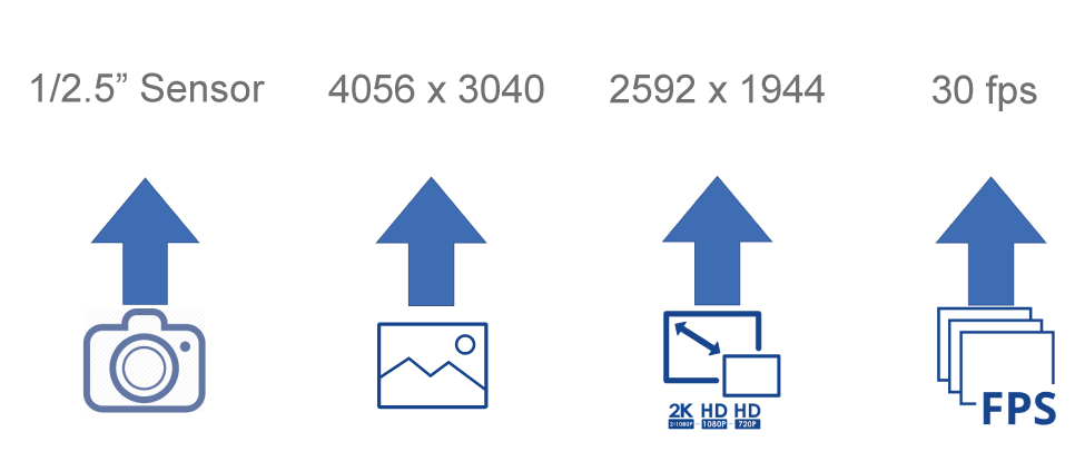
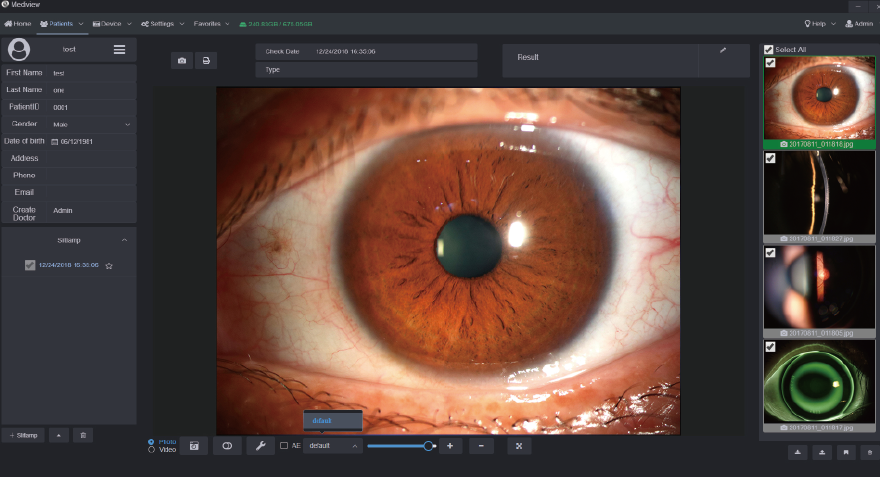
HD Optical System
Optical resolution is up to 2700·N lp/mm
(200 lp/mm), providing more details of the pathologies.
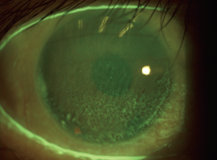
Built-in Yellow Filter
Built-in yellow filter along with cobalt-blue filter increases the contrast of Sodium Fluorescein Staining image.Increase positive rate of early corneal epithelial staining.
The Meibomian Glands Examination
Software Features
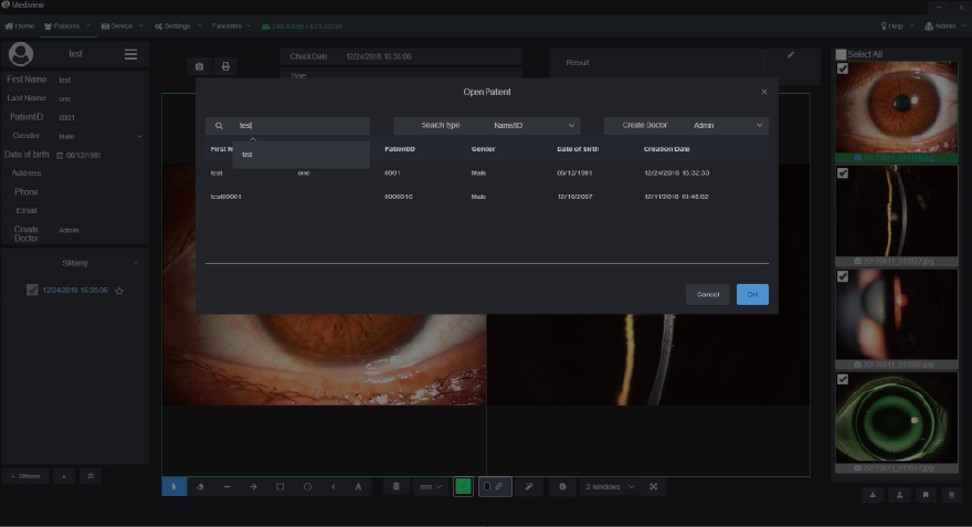
Convenient Patient Management
The patient management system enables clinicians to build and edit patient record,search information by inputting keywords.Clinicians can easily record symptoms and manage the data all the time. The software supports DICOM which makes the images captured by Firefly be easily integrated into hospital’s medical system.
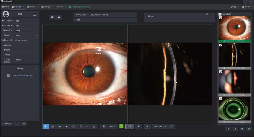
Functional Image Analysis
Clinicians can measure the pathology area with our powerful software tools and change the contrast and brightness of the images. Clinicians can also compare several images at one time to analyze the symptoms and pathology.
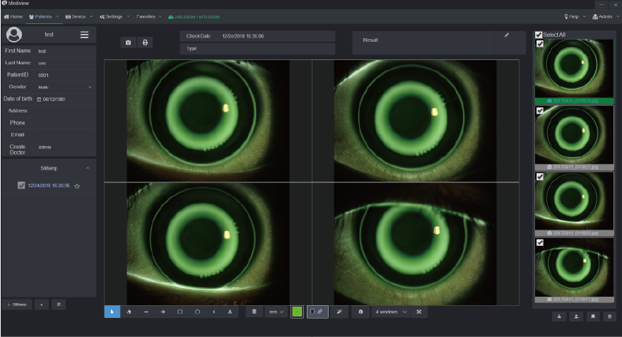
Orthokeratology Lens Fitting Assistance
The optometrists can capture and record high resolution fluorescein images of lens fitting and real-time video without a recording time limit. By comparing the different lens fitting effects, the optometrist can show and educate patients which lens is most suitable for them.
Customized Auto Exposure Value Setting
Clinicians can customize auto exposure values according to the image demand and save as templates for future capturing purpose.
Also, the printing report can be customized according to clinician’s needs.
The Most Effective Tools for Dry Eye
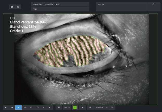
Meibomian Gland Observation
Built-in infrared light source allows the doctor to accurately judge the absence of the meibomian glands.
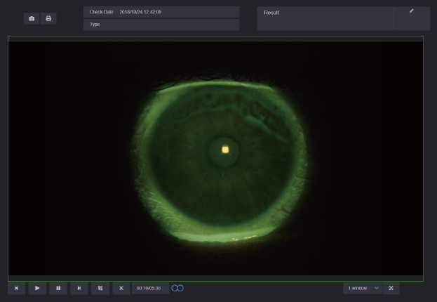
Tear Film Breakup Time
High-performance digital module, doctors can get the tear film breakup time and judge the stability of it by high-resolution video recording.
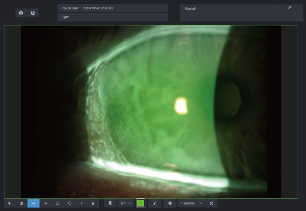
Red Eyes Analysis and Keratopathy Exposure
With a built-in yellow filter, doctors can accurately analyze eye surface damage and inflammation images.
Tear Meniscus Height
Doctors can obtain tear meniscus height by using measuring function in the Mediview software, and effectively evaluate tear
meniscus height.
Related products


